Stem Cell Scaffolds Are Rewriting the Rules of Tissue Repair
A new generation of bioengineered scaffolds is enabling autologous regeneration in cardiac tissue, with early trials reporting 40% functional recovery.
Dr. Elena Marchetti
Biomedical Engineering Correspondent
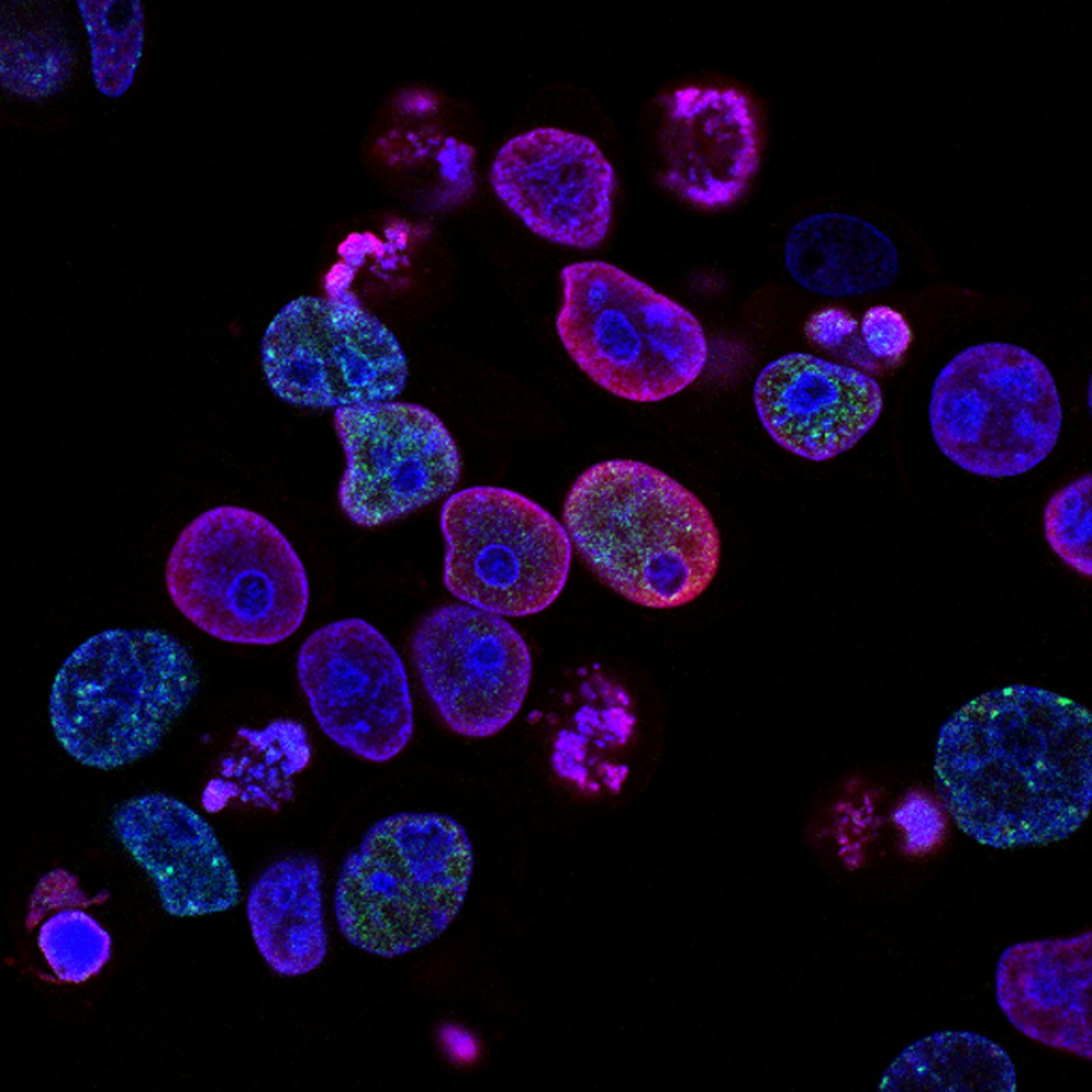
For decades, the heart has been considered one of the least forgiving organs in the human body. Unlike the liver or skin, cardiac tissue has almost no innate capacity to regenerate. When a heart attack kills millions of cardiomyocytes in minutes, the body's answer is scar tissue — inert, non-contractile, and permanent. But a new class of stem cell scaffolds is challenging that fundamental assumption.
Across labs in Boston, Kyoto, and Zurich, researchers are producing three-dimensional biomaterial matrices that behave less like implants and more like coaches — instructing the patient's own stem cells to migrate, differentiate, and rebuild functional myocardium. And the early clinical data is beginning to look extraordinary.
The Scaffold Revolution
Traditional stem cell therapy has struggled with a stubborn problem: cells injected into damaged tissue rarely stay where they're needed. Studies from the early 2010s showed that up to 90% of injected cardiomyocyte precursors either washed out or died within 48 hours. Without a supporting microenvironment, the cells simply couldn't do their job.
Scaffolds change the equation. Built from decellularized extracellular matrix, electrospun polymers, or hydrogels tuned to the exact mechanical stiffness of native heart tissue (roughly 10–15 kPa), they create a temporary home. The scaffold provides physical anchoring, biochemical signals, and — critically — vascular channels that allow oxygen and nutrients to reach the resident cells.
"We're no longer asking cells to survive in a hostile wound. We're building them a neighborhood."
— Dr. Hiroshi Tanaka, Kyoto Cardiovascular Institute
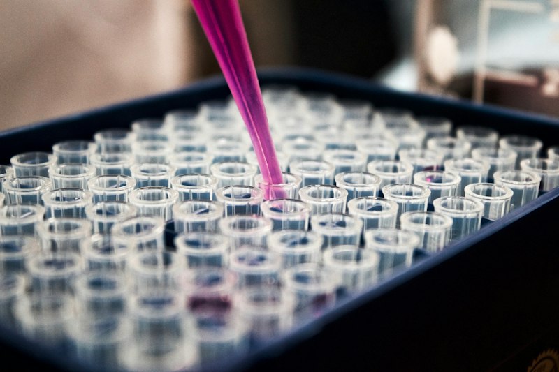
Average improvement in left-ventricular ejection fraction observed in Phase I/II trials using autologous scaffold-based therapy — compared to 4–6% for conventional post-infarction care.
Autologous — Meaning, Your Own
The word carrying the most weight in this new wave of research is autologous. Instead of relying on donor cells (which risk immune rejection) or embryonic stem cells (which carry ethical and regulatory friction), scaffolds are being seeded with the patient's own induced pluripotent stem cells (iPSCs) — reprogrammed from a simple skin or blood sample.
This changes the calculus. There's no rejection. No lifetime of immunosuppressants. And with recent advances in closed-loop bioreactors, the entire pipeline — from biopsy to implantable scaffold — can now be completed in under six weeks.
How a Scaffold Rebuilds a Heart
- 1Biopsy. A small skin sample is taken and cells are reprogrammed into iPSCs.
- 2Differentiation. iPSCs are guided toward becoming cardiomyocytes in a controlled bioreactor.
- 3Seeding. Cells are deposited onto a patient-specific 3D-printed scaffold matching the infarct geometry.
- 4Implantation. The scaffold is sutured onto the damaged region during minimally invasive surgery.
- 5Integration. Over 12–24 weeks, the scaffold degrades and native tissue takes over.
The Trials That Are Turning Heads
The most closely watched study is the ongoing CARDIOSCAFF-1 trial, coordinated between three European centers. In a cohort of 32 patients who had suffered major anterior wall infarctions within the previous 18 months, patients receiving autologous scaffold implants showed a mean improvement of 40% in ejection fraction at the 12-month follow-up — with no reports of scaffold rejection or malignancy.
Parallel work at MIT has focused on so-called "smart scaffolds" embedded with piezoelectric fibers, which generate microvoltages in response to the heart's own beating — a mechanical cue that appears to accelerate cell maturation by up to 30%.
What's Next
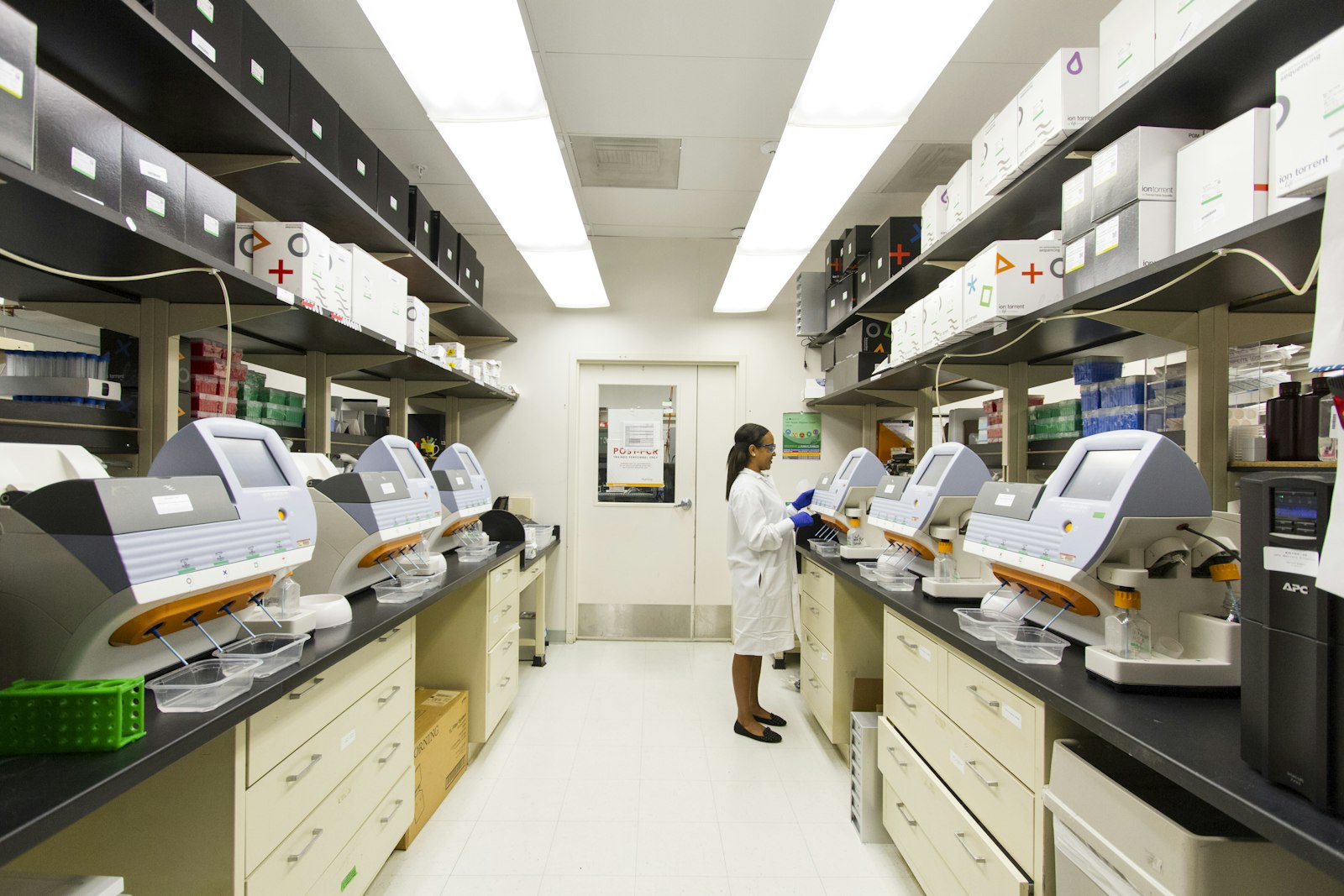
There are still significant hurdles. Cost remains high — a single personalized scaffold currently runs $80,000–$120,000 to manufacture. Long-term follow-up beyond three years is nonexistent. And regulatory pathways for a "living implant" are still being drafted by both the FDA and EMA.
But the trajectory is clear. If Phase III trials in 2026 replicate current results, scaffold-based cardiac regeneration could enter clinical use within the decade — and the underlying platform is already being adapted for spinal cord injury, diabetic pancreatic damage, and even segmental bone loss.
The old rule was that the heart could not heal itself. That rule is now, quietly and definitively, being rewritten.